Vascular Ultrasound Services in
At Minimally Invasive Specialists of Texas, we offer comprehensive vascular ultrasound services — a safe, non-invasive, radiation-free imaging technique that uses high-frequency sound waves to evaluate the health and function of your arteries and veins in real time. From detecting blood clots and peripheral arterial disease to mapping varicose veins and assessing venous insufficiency, our vascular ultrasounds are performed by skilled technicians and interpreted by our interventional radiology specialists to ensure accurate diagnoses and guide the most effective treatment plans. Call (832) 583-2246 - Pasadena Office , (832) 583-2246 - Baytown Office or (832) 583-2246 - Clear Lake Office to schedule your study in .
What Is a Vascular Ultrasound?
A vascular ultrasound — also known as a duplex ultrasound or Doppler ultrasound — is a non-invasive diagnostic imaging study that uses high-frequency sound waves to create detailed, real-time images of the body’s blood vessels. Unlike CT or X-ray imaging, vascular ultrasound uses no radiation, requires no intravenous contrast dye in most cases, and carries no known health risks, making it safe for patients of all ages including those with kidney disease, contrast allergies, or pregnancy.
The term “duplex” ultrasound refers to the combination of two imaging modes used simultaneously: B-mode imaging, which produces structural grayscale images of the vessel walls and surrounding tissue, and Doppler imaging, which measures the speed and direction of blood flow within the vessel in real time. Together, these two capabilities allow our team at Minimally Invasive Specialists of Texas to not only see the anatomy of a blood vessel but also assess how well blood is actually flowing through it — detecting blockages, narrowing, clots, and valvular dysfunction that would otherwise be invisible. Call (832) 583-2246 - Pasadena Office , (832) 583-2246 - Baytown Office or (832) 583-2246 - Clear Lake Office to schedule your vascular ultrasound in .
What Conditions Can Vascular Ultrasound Diagnose?
Vascular ultrasound is one of the most versatile and widely used diagnostic tools in vascular medicine, providing essential information for the evaluation and management of a broad range of conditions. At Minimally Invasive Specialists of Texas, our vascular ultrasound services are used to diagnose and monitor:
- Deep Venous Thrombosis (DVT): Vascular ultrasound is the primary diagnostic test for blood clots in the deep veins of the legs, thighs, or pelvis. It can confirm the presence, location, and extent of a clot and guide decisions about anticoagulation or interventional treatment. Learn more on our DVT page.
- Peripheral Arterial Disease (PAD): Duplex arterial ultrasound of the lower extremities maps the arteries of the legs, identifies sites of stenosis or occlusion, and quantifies the degree of flow reduction — providing essential information for treatment planning. Learn more on our PAD page.
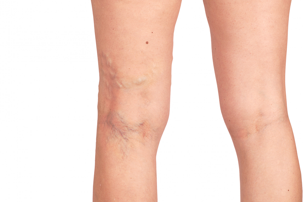
- Venous Insufficiency and Varicose Veins: A standing duplex ultrasound of the legs evaluates the function of the venous valves and identifies the specific sites of venous reflux (backward blood flow) that cause varicose veins and chronic venous disease — a critical step before any vein treatment. Learn more on our chronic venous disease page.
- Pelvic Venous Insufficiency / Pelvic Congestion Syndrome: Pelvic and transvaginal Doppler ultrasound can identify dilated ovarian and pelvic veins with retrograde flow — key findings in the diagnosis of pelvic congestion syndrome.
- Carotid Artery Disease: Carotid duplex ultrasound evaluates the carotid arteries in the neck for plaque buildup, stenosis, and flow abnormalities that increase the risk of stroke.
- Abdominal Aortic Aneurysm (AAA): Ultrasound is used to screen for and monitor enlargement of the abdominal aorta, enabling timely intervention before rupture risk increases.
- Renal Artery Stenosis: Duplex ultrasound of the renal arteries can detect significant narrowing that contributes to difficult-to-control hypertension or kidney dysfunction.
- Dialysis Access Surveillance: Regular ultrasound evaluation of arteriovenous fistulas and grafts used for hemodialysis detects stenosis and flow problems before they cause access failure. Learn more on our dialysis access page.
- Pseudoaneurysms and Arteriovenous Fistulas: Post-procedural complications involving abnormal connections or pulsatile collections adjacent to blood vessels can be precisely characterized with Doppler ultrasound.
- Venous Mapping: Before surgical bypass or vein harvest procedures, ultrasound maps the superficial veins to assess their suitability for use as bypass conduits.
Types of Vascular Ultrasound Studies We Perform
At Minimally Invasive Specialists of Texas, our vascular laboratory offers a comprehensive menu of ultrasound studies tailored to the specific clinical question being evaluated:
- Lower Extremity Venous Duplex (DVT Study): Evaluates the deep veins of the leg from the groin to the calf for the presence of blood clots. Performed with the patient supine and in various positions to optimize visualization of all venous segments.
- Lower Extremity Arterial Duplex: Maps the arteries of the leg from the aorto-iliac segment to the tibial vessels at the ankle, identifying the location and severity of arterial stenosis or occlusion in patients with symptoms of PAD.
- Venous Reflux Study (Venous Insufficiency Study): A standing duplex examination that assesses venous valve function throughout the superficial and deep venous systems of the legs, identifying pathological reflux responsible for varicose veins and chronic venous symptoms.
- Carotid Duplex Ultrasound: Evaluates the common, internal, and external carotid arteries bilaterally for plaque, stenosis, and flow abnormalities associated with stroke risk.
- Abdominal Aortic Ultrasound: Measures the diameter of the abdominal aorta to screen for or monitor an abdominal aortic aneurysm.
- Renal Artery Duplex: Assesses blood flow in the renal arteries and intrarenal vessels to detect stenosis contributing to renovascular hypertension or ischemic nephropathy.
- Dialysis Access Duplex (Fistula / Graft Surveillance): Evaluates the entire dialysis access circuit for flow volumes, stenosis, and other abnormalities that may predict access failure.
- Mesenteric Artery Duplex: Assesses blood flow in the superior and inferior mesenteric arteries in patients with symptoms of chronic mesenteric ischemia.
What to Expect During a Vascular Ultrasound at Minimally Invasive Specialists of Texas
Vascular ultrasound examinations at Minimally Invasive Specialists of Texas are safe, comfortable, and require no special preparation in most cases. Here is what patients can expect:
- No radiation, no needles, no contrast: The entire examination uses only sound waves transmitted through a handheld probe (transducer) placed gently against the skin. There is no radiation, no injections, and no contrast dye required for most studies.
- Gel application: A water-based gel is applied to the skin overlying the area being examined to ensure good acoustic contact between the probe and the skin.
- Real-time imaging: The sonographer moves the transducer over the skin to capture images of the target vessels from multiple angles. For venous reflux studies, you may be asked to stand or change positions during the examination.
- Duration: Most vascular ultrasound studies take 30 to 60 minutes depending on the number of vessels being evaluated and the clinical question being answered.
- Interpretation by specialists: All studies performed at Minimally Invasive Specialists of Texas are interpreted by our experienced interventional radiology physicians, ensuring accurate and clinically relevant reporting that directly guides your treatment plan.
Results are typically available promptly and communicated to your referring physician. If your ultrasound identifies a condition requiring interventional treatment, our team at Minimally Invasive Specialists of Texas is uniquely positioned to provide seamless, same-practice care — from diagnosis through treatment. Call (832) 583-2246 - Pasadena Office , (832) 583-2246 - Baytown Office or (832) 583-2246 - Clear Lake Office to schedule your vascular ultrasound in .
Why Vascular Ultrasound Is Essential for Treatment Planning
For many of the conditions treated at Minimally Invasive Specialists of Texas, a vascular ultrasound is not simply a diagnostic nicety — it is an essential prerequisite to safe and effective treatment. Before performing vein ablation for varicose veins, our interventional radiologists use venous duplex mapping to identify the precise sites of reflux, determine which veins require treatment, and plan the safest access points. Before angioplasty or stenting for PAD, arterial duplex data helps confirm the lesion location and severity. Before dialysis access intervention, surveillance ultrasound identifies stenosis before it progresses to thrombosis and access failure.
By performing vascular ultrasound in-house at Minimally Invasive Specialists of Texas, we ensure that the physician interpreting your images is the same specialist who will be treating your condition — enabling a level of diagnostic-therapeutic integration that leads to better outcomes. Our full range of vascular conditions treated can be found on our services page. Call us at (832) 583-2246 - Pasadena Office , (832) 583-2246 - Baytown Office or (832) 583-2246 - Clear Lake Office to get started in .
Frequently Asked Questions About Vascular Ultrasound
Is a vascular ultrasound painful or uncomfortable?
No — vascular ultrasound is a completely painless and non-invasive procedure. The examination involves only a handheld probe placed gently against the skin with a small amount of water-based gel to improve image quality. There are no needles, no injections, no radiation, and no discomfort associated with the procedure itself. Some patients may experience mild pressure when the probe is pressed firmly over deeper vessels, but this is not painful. If you have concerns about the examination, our team at Minimally Invasive Specialists of Texas is happy to walk you through what to expect before you arrive — call (832) 583-2246 - Pasadena Office , (832) 583-2246 - Baytown Office or (832) 583-2246 - Clear Lake Office .
Do I need to prepare for a vascular ultrasound?
Preparation requirements vary depending on the type of study being performed. Most lower extremity venous and arterial ultrasounds require no special preparation — you can eat, drink, and take your regular medications as normal. Abdominal aortic and mesenteric artery studies typically require fasting for several hours beforehand to minimize bowel gas that can interfere with imaging. Our team at Minimally Invasive Specialists of Texas will provide you with specific preparation instructions when you schedule your appointment. Call (832) 583-2246 - Pasadena Office , (832) 583-2246 - Baytown Office or (832) 583-2246 - Clear Lake Office with any questions in .
How long does a vascular ultrasound take?
The duration of a vascular ultrasound examination at Minimally Invasive Specialists of Texas depends on the type and extent of the study. Most single-region studies — such as a lower extremity DVT evaluation or a carotid duplex — take approximately 30 to 45 minutes. More comprehensive studies evaluating multiple vessel segments, such as a bilateral arterial duplex of the legs or a full venous reflux mapping, may take 45 to 60 minutes or longer. Our scheduling team will give you an estimated time when you book your appointment. Call (832) 583-2246 - Pasadena Office , (832) 583-2246 - Baytown Office or (832) 583-2246 - Clear Lake Office to schedule in .
What is the difference between a vascular ultrasound and a regular ultrasound?
A standard ultrasound creates structural images of organs and soft tissues — such as the liver, kidneys, or uterus — to assess their anatomy. A vascular (duplex) ultrasound is specifically designed to evaluate blood vessels, combining structural B-mode imaging of the vessel walls with Doppler technology that measures the speed and direction of blood flow in real time. This dual capability allows vascular ultrasound to detect not just anatomical abnormalities such as plaque or clots, but also functional problems such as valve incompetence, turbulent flow, or stenosis that would be invisible to a standard structural ultrasound.
Will my insurance cover a vascular ultrasound at Minimally Invasive Specialists of Texas?
Vascular ultrasound studies are covered by Medicare, Medicaid, and most private insurance plans when they are ordered for a medically indicated reason — such as evaluation of leg swelling, suspected DVT, claudication symptoms, or varicose vein mapping before treatment. Coverage and prior authorization requirements vary by insurer and specific study type. Our team at Minimally Invasive Specialists of Texas will help verify your benefits and ensure the study is ordered and documented appropriately to support insurance coverage. Call (832) 583-2246 - Pasadena Office , (832) 583-2246 - Baytown Office or (832) 583-2246 - Clear Lake Office for assistance with your coverage questions in .
Do I need a referral from my doctor to get a vascular ultrasound at Minimally Invasive Specialists of Texas?
In most cases, a referral or order from your primary care physician or a specialist is required to perform a vascular ultrasound, both for insurance purposes and to ensure the correct study is ordered based on your clinical symptoms. However, we encourage you to call our office at (832) 583-2246 - Pasadena Office , (832) 583-2246 - Baytown Office or (832) 583-2246 - Clear Lake Office — our team can help coordinate the referral process with your physician and ensure you receive the appropriate study in a timely manner. If your ultrasound identifies a condition requiring treatment, our interventional radiology team is ready to guide next steps.
How quickly will I receive my vascular ultrasound results?
At Minimally Invasive Specialists of Texas, all vascular ultrasound studies are interpreted promptly by our interventional radiology specialists. In most cases, a formal report is generated and transmitted to your referring physician within one business day of the examination. If the study reveals an urgent finding — such as an acute DVT or critical arterial occlusion — our team will communicate results to your physician on the same day. If you have questions about your results or what they mean for your care, call us at (832) 583-2246 - Pasadena Office , (832) 583-2246 - Baytown Office or (832) 583-2246 - Clear Lake Office and our team in will be happy to assist.